What Is an Esophageal Ulcer?
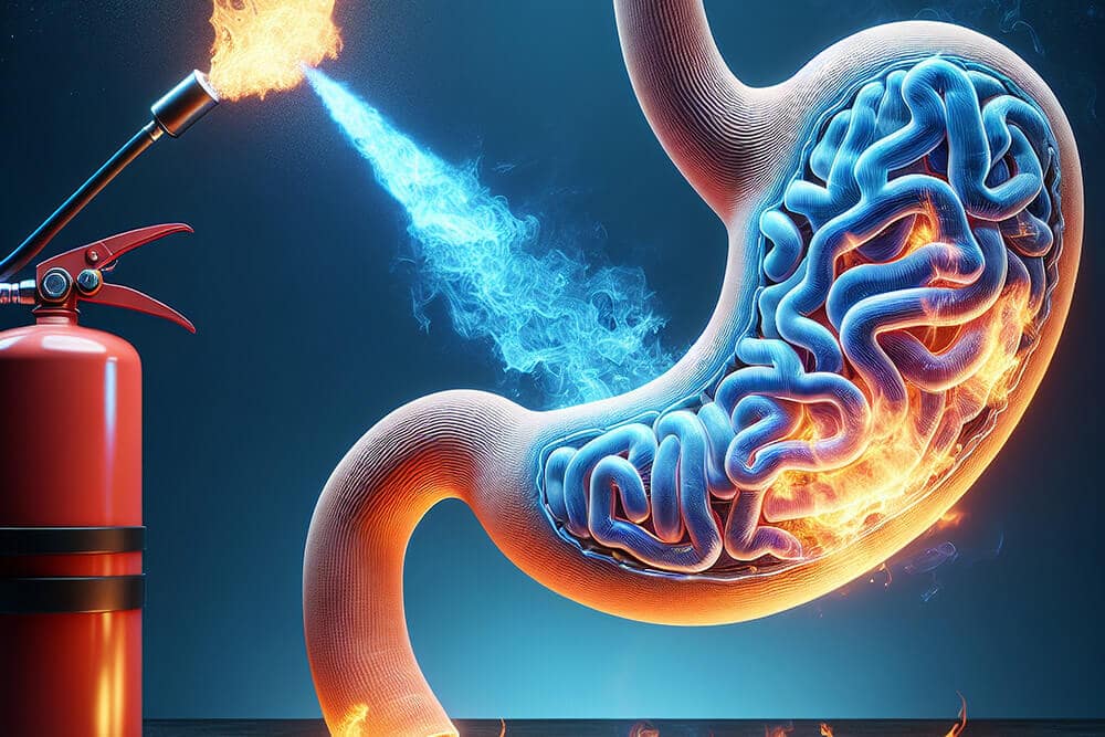
Esophageal ulcer refers to a rupture or defect in the lining of the tube, which pushes food in your mouth to your stomach, known as the esophagus. The stomach acid or any other irritant attacks the protective lining of the stomach leading to its development. Unattended ulcers may also bleed, scar and constrict the esophagus, although they can be treated with numerous weeks of medical support.
Common Causes and Risk Factors
- Gastroesophageal reflux disease (GERD) -chronic acid reflux
- Infections -fungal (Candida) or viral (herpes, CMV)
- Medications -NSAIDs, aspirin, certain antibiotics
- Smoking -tobacco weakens and inflames esophageal tissue
- Alcohol use -irritates and thins the mucosal lining
- Hiatal hernia -allows stomach acid to back up into the esophagus
Signs and Symptoms
- Burning or gnawing chest pain that may worsen with eating
- Pain or discomfort when swallowing (odynophagia)
- Chest pain behind the breastbone
- Nausea or episodes of vomiting
- Unintended weight loss or decreased appetite
- Signs of bleeding -dark stools or anemia (advanced cases)
Expert Treatment for Esophageal Ulcer by Dr. Bharat Pothuri
Dr. Pothuri uses a step-by-step approach:
Medical History and Physical Exam
He questions whether or not you have pain in your chest, or epigastric area or heartburn, have trouble with swallowing, how long have your had the pain, have you been taking any medications (NSAIDs, antibiotics), do you use alcohol and tobacco, and have you lost any weight. He will even feel your abdomen and look whether it is tender or not.
Blood Tests
Anemia due to chronic hemorrhage could be seen by a complete blood count (CBC). H. pylori antibodies or antigen tests will show infection which will commonly lead to the development of ulcers.
Imaging and Functional Studies
- Barium Swallow X-Ray: Evaluates anatomy, detects strictures, hiatal hernia, or motility disorders.
- Esophageal pH Monitoring: Measures acid exposure to confirm reflux as a cause of ulceration and pain.
- Esophageal Manometry: Assesses muscle contractions and sphincter function if motility issues are suspected.
Upper Endoscopy (EGD)
A small, bony scope is introduced into the esophagus to enable direct observation of the sores, ulcers, or inflammation. To eliminate yeast, viral, dysplasia/cancer, biopsies, and tissue cultures can be obtained.
Advanced Testing (if needed)
Under certain circumstances, endoscopic ultrasound or CT scan is used to measure the extent of involvement of deep tissue, eliminate complications, or inform additional treatment.

Frequently Asked Questions
Where is the ICD-10 code of an esophageal ulcer?
K22.1 is the ICD-10 code used in medical records and insurance billing by healthcare providers.
What is the recovery time for an esophageal ulcer?
With proper treatment, most esophageal ulcers heal within 4–8 weeks.
Can an esophageal ulcer develop into cancer?
Chronic esophageal ulcers may raise concern, but progression to cancer is uncommon. Dr. Pothuri monitors patients closely with follow-up evaluations.
How is an esophageal ulcer managed?
Management includes eating soft foods, avoiding spicy or acidic meals, quitting smoking, limiting alcohol, and taking prescribed medications as directed by your doctor.
When should I seek emergency care for chest pain?
If chest pain is severe, sudden, or radiates to other areas (e.g., arm, jaw, back), it may indicate a medical emergency. Call 911 or go to the nearest emergency room immediately.
Is endoscopy always necessary?
Endoscopy provides the most accurate visual assessment and allows for biopsy if needed. Dr. Pothuri will recommend endoscopy based on your symptoms and risk factors.